Your Shopping Cart is empty.


Download sample
File Details
Published: 2024-09-03 09:29:11.236659 Category: Graphic Resources Type: Illustration Model release: No
Share
       

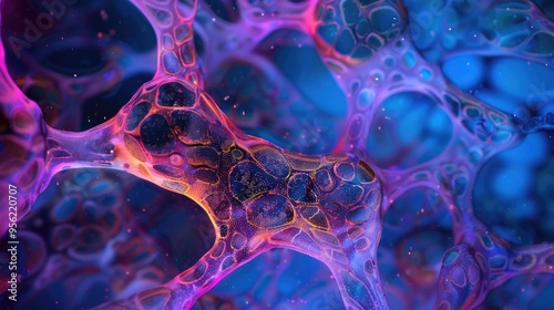
Low magnification light microscope micrograph of cross sectioned myelinated nerve fascicles stained with osmium tetroxyde to show the myelin sheath of nerve fibers

Contributor: Gular
ID : 956220707

TitleFilesize
Illustration7280x4080


Buy on Adobe Stock

Keywords
microscope, cross section, nerve, stained, osmium, sheath, fiber, low, magnification, light, microscopy, histology, tissue, cell, structure, nervous, system, peripheral, central, laboratory, science, research, imaging, cellular, detail, visualisation, electron, study, diagnostic, medicals, biology, physiology, nerve cell, staining, preparation, quality